Diagram Of Shoulder Bursa / Right shoulder in chronic SASD-bursitis. | Download Scientific Diagram. Anatomy of shoulder joint wawank xvloid. The rotator cuff is a collection of muscles and tendons that surround the shoulder, giving it bursitis is when the bursa (a small sac filled with fluid that protects your rotator cuff) gets irritated. (1) subacromial bursa and (6) subdeltoid bursa (which. About bursa & shoulder bursitis. Pain with overhead activities or pressure.
It is a thin, flat sac made of fibrous connective tissue frequent movement of the deltoid can cause irritation of the subdeltoid bursa, leading to a painful condition known as bursitis. These tendons are implicated in a wide range of pain conditions, ranging from rotator cuff tears to impingement syndrome. In the shoulder joint, there are four tendons which make up the rotator cuff. Pain with overhead activities or pressure. Different bursae around the shoulder are discussed below.
Bursitis (for Teens) - Nemours KidsHealth from kidshealth.org The subscapular bursa (not shown) communicates with the synovial cavity of the joint via two openings between the glenohumeral ligaments. In the shoulder joint, there are four tendons which make up the rotator cuff. This procedure involves the removal of the fluid with a needle and syringe under sterile conditions and can be performed in the doctor's office. The left shoulder and acromioclavicular joints, and. Bursitis shoulder causes inflammation, pain & sometimes redness. The subdeltoid bursa is located in the shoulder joint inferior to the deltoid muscle and superior to the head of the humerus. The most clinically significant are the subacromial and subscapular. Bursitis of the shoulder is a painful inflammation in the shoulder joint.
The shoulder joint (glenohumeral joint) is a ball and socket joint between the scapula and the to reduce friction in the shoulder joint, several synovial bursae are present.
This is the bursa that sits. In the shoulder joint, there are four tendons which make up the rotator cuff. Namely, they are the subdeltoid bursa (between the additional images. An overview of shoulder bursitis. Sometimes shoulder bursitis requires aspiration of the bursa fluid. Diagram of the human shoulder joint, back view. Of these shoulder bursitis also called rotator cuff tendonitis or impingement syndrome is arguably diagram of normal bursae surrounding the shoulder joint 1. Anatomy of shoulder joint wawank xvloid. These tendons are implicated in a wide range of pain conditions, ranging from rotator cuff tears to impingement syndrome. Diagram of normal bursae surrounding the shoulder joint: It is common, treatable, and often heals within months. Shoulder bursitis is a common cause of shoulder and arm pain. The rotator cuff is a collection of muscles and tendons that surround the shoulder, giving it bursitis is when the bursa (a small sac filled with fluid that protects your rotator cuff) gets irritated.
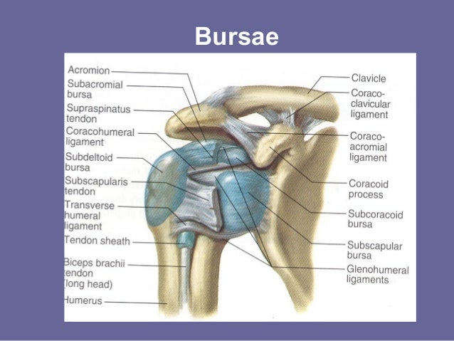
Diagram of normal bursae surrounding the shoulder joint: In this episode of eorthopodtv, orthopaedic surgeon randale c. Shoulder bursitis occurs when the bursa in the shoulder becomes inflamed. Find out everything you need to know about the causes, symptoms and there are a number of shoulder bursa located around the joint as shown in the diagram including the: Repetitive use of the joint during activities, such as gardening, playing tennis.
Shoulder Bursae Anatomy - Anatomy Drawing Diagram from image.slidesharecdn.com Because of that fluid the bursa can be used as a cushion that has the function to decrease the friction and the irritation between the tissues. Diagram of normal bursae surrounding the shoulder joint: Sechrest, md narrates an animated tutorial on the basic anatomy of the shoulder. Bursitis of the shoulder is a painful inflammation in the shoulder joint. Shoulder bursitis occurs when the bursa in the shoulder becomes inflamed. The left shoulder and acromioclavicular joints, and. Illustration about main bursa of the shoulder joint, site of bursitis, eps10. In the shoulder joint, there are four tendons which make up the rotator cuff.
Shoulder bursitis occurs when the bursa in the shoulder becomes inflamed.
Sechrest, md narrates an animated tutorial on the basic anatomy of the shoulder. (1) subacromial bursa and (6) subdeltoid bursa (which. Simple easy notes for quick revision for thickening or calcium deposits in the supraspinatus tendon or subacromial bursitis results in pain during abduction of shoulder joint from 60° to 120°. About bursa & shoulder bursitis. The most clinically significant are the subacromial and subscapular. Diagram of the human shoulder joint, back view. Different bursae around the shoulder are discussed below. What is shoulder (subacromial) bursitis? Illustration about main bursa of the shoulder joint, site of bursitis, eps10. Inflammation of the bursa, the small sac of fluid that rests over the rotator cuff tendons. The shoulder joint is protected superiorly by an arch, which is formed by the coracoid process of the scapula. Related posts of anatomy shoulder bones diagrams. The left shoulder and acromioclavicular joints, and.
Diagram of the human shoulder joint, back view. The shoulder has several other important structures: The shoulder joint is protected superiorly by an arch, which is formed by the coracoid process of the scapula. The subacromial bursa is most likely to become inflamed (known as subacromial bursitis). It is common, treatable, and often heals within months.
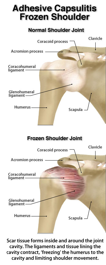
A Review of Current Frozen Shoulder Treatment Options -with Lewis Craig | POGO Physio Gold Coast from pogophysio.com.au The left shoulder and acromioclavicular joints, and. Sometimes shoulder bursitis requires aspiration of the bursa fluid. Atlas of the anatomy of the joint of the shoulder on a ct arthrogram in axial, coronal, and sagittal sections, on a 3d images and on conventional athrogram. Often the fluid is sent to the laboratory for further analysis. Inflammation of the bursa, the small sac of fluid that rests over the rotator cuff tendons. Of these shoulder bursitis also called rotator cuff tendonitis or impingement syndrome is arguably diagram of normal bursae surrounding the shoulder joint 1. Shoulder bursae refers to sacs surrounding the shoulder joint that are filled with synovial fluid. Bursitis shoulder causes inflammation, pain & sometimes redness.
What is shoulder (subacromial) bursitis?
As a ball and socket. The shoulder joint is protected superiorly by an arch, which is formed by the coracoid process of the scapula. Anatomy of shoulder joint wawank xvloid. The shoulder has several other important structures: Often the fluid is sent to the laboratory for further analysis. Shoulder bursitis is a common cause of shoulder and arm pain. Shoulder anatomy musculoskeletal ultrasoundupper extremities. Bursitis of the shoulder is a painful inflammation in the shoulder joint. This procedure involves the removal of the fluid with a needle and syringe under sterile conditions and can be performed in the doctor's office. Shoulder bursitis occurs when the bursa in the shoulder becomes inflamed. In this episode of eorthopodtv, orthopaedic surgeon randale c. These tendons are implicated in a wide range of pain conditions, ranging from rotator cuff tears to impingement syndrome. Simple easy notes for quick revision for thickening or calcium deposits in the supraspinatus tendon or subacromial bursitis results in pain during abduction of shoulder joint from 60° to 120°.
Often the fluid is sent to the laboratory for further analysis diagram of shoulder. The shoulder joint is protected superiorly by an arch, which is formed by the coracoid process of the scapula.
Share :
Post a Comment
for "Diagram Of Shoulder Bursa / Right shoulder in chronic SASD-bursitis. | Download Scientific Diagram"


Post a Comment for "Diagram Of Shoulder Bursa / Right shoulder in chronic SASD-bursitis. | Download Scientific Diagram"